|
People often
wonder what a comprehensive eye examination involves, what it tells the
physician, and how the physician uses the results from an eye exam to devise
a treatment program.
The first thing
an ophthalmologist or optometrist checks are the eye's vital signs: visual
acuity, eye pressure, and visual fields.
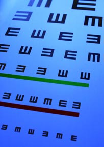
Visual acuity:
Visual acuity is measured on an eye chart and recorded using numbers like
20/20 or 20/50. Looking through a single pinhole or multiple pinholes can
give an estimate of the best vision that could be achieved if the lenses in
a pair of eyeglasses were further optimized. This is an easy and useful
method to assess whether a change in a lens prescription would be
beneficial. Another technique for testing best visual acuity is called
refraction. Here, different lenses are placed in front of the eyes to
determine which would improve vision.
Eye pressure:
This is measured using a special device called a tonometer. One form of this
instrument is about the size of a fountain pen and is applied to the cornea
for a few seconds to measure the pressure of the eye. This test screens for
glaucoma and other eye health problems.
Visual fields:
A test of visual fields measures peripheral vision. Macular degeneration
affects central vision and usually spares peripheral vision.
The
doctor will also take a patient's medical history. A complete examination is
multi-stepped and includes gathering information about past medical and
ocular disorders, medications, nutrition, allergies, history of smoking and
alcohol use, and family medical problems. Then comes the actual eye exam.
Examination of
the front portion of the eye
The front of the eye includes the parts we see in the mirror plus the lens
which lies behind the pupil.
Motility of the
eye:
The doctor checks whether the eyes move freely and in alignment. The
alignment is very important in order to avoid double vision.
Eyelids:
The eyelids are checked for evidence of infection or malfunction.
Conjunctiva:
The conjunctiva is a thin layer of cells covering the portion of the eyeball
between the lids. Inflammation and redness of the conjunctiva can be a sign
of infection or allergy.
Tear film:
As we age, the tear film often becomes less capable of keeping the surface
of the eye properly lubricated. This is called dry eye and is not related to
the dry form of macular degeneration.
Cornea:
The cornea is the clear front surface of the eye that is responsible for
most of the eye's focusing power. In refractive surgery, lasers are used to
change the curvature of the surface of the cornea so that eyeglasses or
contact lenses are no longer needed. The doctor looks for evidence of
corneal irritation, inflammation, and infection.
Lens:
The other main component for focusing images is the lens. When the lens
becomes cloudy it is called a cataract. A cataract that becomes cloudy
enough to significantly impair vision can be removed and replaced with an
artificial lens (called an implant). The lens is examined to determine if a
cataract is present and to judge its severity. |