|
What is a cataract?
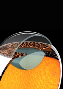
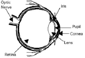
The lens is made mostly of
water and protein. The protein is arranged to let light pass through and
focus on the retina. Sometimes some of the protein clumps together. This can
start to cloud small areas of the lens, blocking some light from reaching
the retina and interfering with vision. This is a cataract.
In its early stages, a
cataract may not cause a problem. The cloudiness may affect only a small
part of the lens. However, over time, the cataract may grow larger and cloud
more of the lens, making it harder to see. Because less light reaches the
retina, your vision may become dull and blurry. A cataract won't spread from
one eye to the other, although many people develop cataracts in both eyes.
Although researchers are
learning more about cataracts, no one knows for sure what causes them.
Scientists think there may be several causes, including smoking, diabetes,
and excessive exposure to sunlight.
What are
the symptoms?
The most common symptoms of
a cataract are:
● Cloudy or blurry vision.
● Problems with light.
These can include headlights that seem too bright at night; glare from lamps
or very bright sunlight; or a halo around lights.
● Colors that seem faded.
● Poor night vision.
● Double or multiple vision
(this symptom often goes away as the cataract grows).
● Frequent changes in your
eyeglasses or contact lenses.
These symptoms can also be
a sign of other eye problems. If you have any of these symptoms, check with
your eye care professional.
When a cataract is small,
you may not notice any changes in your vision. Cataracts tend to grow
slowly, so vision gets worse gradually. Some people with a cataract find
that their close-up vision suddenly improves, but this is temporary. Vision
is likely to get worse again as the cataract grows.
What are
the different types of cataract?
Age-related
cataract:
Most cataracts are related to aging.
Congenital
cataract:
Some babies are born with cataracts or develop them in childhood, often in
both eyes. These cataracts may not affect vision. If they do, they may need
to be removed.
Secondary
cataract:
Cataracts are more likely to develop in people who have certain other health
problems, such as diabetes. Also, cataracts are sometimes linked to steroid
use.
Traumatic
cataract:
Cataracts can develop soon after an eye injury, or years later.
How is a
cataract detected?
To detect a
cataract, an eye care professional examines the lens. A comprehensive eye
examination usually includes:
Visual
acuity test: This eye chart test measures how well you see at various
distances.
Pupil
dilation:
The pupil is widened with eyedrops to allow your eye care professional to
see more of the lens and retina and look for other eye problems.
Tonometry:
This is a standard test to measure fluid pressure inside the eye. Increased
pressure may be a sign of glaucoma.
How is it
treated?
For an early
cataract, vision may improve by using different eyeglasses, magnifying
lenses, or stronger lighting. If these measures don't help, surgery is the
only effective treatment. This treatment involves removing the cloudy lens
and replacing it with a substitute lens.
A cataract
needs to be removed only when vision loss interferes with your everyday
activities, such as driving, reading, or watching TV.
Is
cataract surgery effective?
Cataract
removal is one of the most common operations performed in the U.S. today. It
is also one of the safest and most effective. In about 90 percent of cases,
people who have cataract surgery have better vision afterward.
How is a
cataract removed?
There are two
primary ways to remove a cataract. Your doctor can explain the differences
and help determine which is best for you:
Phacoemulsification, or phaco.
Your doctor makes a small incision on the side of the cornea, the clear,
dome-shaped surface that covers the front of the eye. The doctor then
inserts a tiny probe into the eye. This device emits ultrasound waves that
soften and break up the cloudy center of the lens so it can be removed by
suction. Most cataract surgery today is done by phaco, which is also called
small incision cataract surgery.
Extracapsular surgery.
Your doctor makes a slightly longer incision on the side of the cornea and
removes the hard center of the lens. The remainder of the lens is then
removed by suction.
In most
cataract surgeries, the removed lens is replaced by an intraocular lens (IOL).
An IOL is a clear, artificial lens that requires no care and becomes a
permanent part of your eye. With an IOL, you'll have improved vision because
light will be able to pass through it to the retina. Also, you won't feel or
see the new lens.
Some people
cannot have an IOL. They may have problems during surgery, or maybe they
have another eye disease. For these people, a soft contact lens may be
suggested. For others, glasses that provide powerful magnification may be
better.
What
happens before surgery?
A week or two before
surgery, your eye care professional will do some tests. These may include
tests to measure the curve of the cornea and the size and shape of the eye.
For patients who will receive an IOL, this information helps your doctor
choose the right type of IOL. Also, doctors may ask you not to eat or drink
anything after midnight the morning of your surgery.
What
happens during surgery?
When you enter the hospital
or clinic, you will be given eye drops to dilate the pupil. The area around
your eye will be washed and cleansed.
The operation usually lasts
less than 1 hour and is almost painless. Many people choose to stay awake
during surgery, while others may need to be put to sleep for a short time.
If you are awake, you will have an anesthetic to numb the nerves in and
around your eye.
After the operation, a
patch will be placed over your eye and you will rest for a while. You will
be watched by your medical team to see if there are any problems, such as
bleeding. Most people who have cataract surgery can go home the same day.
Since you will not be able to drive, make sure you make arrangements for a
ride.
What
happens after surgery?
It's normal to
feel itching and mild discomfort for a while after cataract surgery. Some
fluid discharge is also common, and your eye may be sensitive to light and
touch. If you have discomfort, your eye care professional may suggest a pain
reliever every 4-6 hours. After 1-2 days, even moderate discomfort should
disappear. In most cases, healing will take about 6 weeks.
After surgery,
your doctor will schedule exams to check on your progress. For a few days
after surgery, you may take eyedrops or pills to help healing and control
the pressure inside your eye. Ask your doctor how to use your medications,
when to take them, and what effects they can have. You will also need to
wear an eye shield or eyeglasses to help protect the eye. Avoid rubbing or
pressing on your eye.
Problems after
surgery are rare, but they can occur. These can include infection, bleeding,
inflammation (pain, redness, swelling), loss of vision, or light flashes.
With prompt medical attention, these problems usually can be treated
successfully.
When you are
home, try not to bend or lift heavy objects. Bending increases pressure in
the eye. You can walk, climb stairs, and do light household chores.
When will
my vision be normal again?
You can quickly return to
many everyday activities, but your vision may be blurry. The healing eye
needs time to adjust so that it can focus properly with the other eye,
especially if the other eye has a cataract. Ask your doctor when you can
resume driving.
If you just received an IOL,
you may notice that colors are very bright or have a blue tinge. Also, if
you've been in bright sunlight, everything may be reddish for a few hours.
If you see these color tinges, it is because your lens is clear and no
longer cloudy. Within a few months after receiving an IOL, these colors
should go away. And when you have healed, you will probably need new
glasses. |